
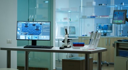
Breast-conserving surgery aims to remove tumors while preserving as much healthy breast tissue as possible, ensuring optimal aesthetic outcomes that are critical for a patient’s quality of life. To achieve this objective, precise location of the tumor is necessary during surgery, which is normally performed with the patient lying face-up (supine position). To plan the procedure, surgeons rely on medical imaging technologies that often provide limited perspectives due to the conditions under which the images are captured. For instance, MRI scans are performed with the patient lying face down (prone position), causing the breast to compress and deform, while 3D scans capture the natural shape of the breast in the upright position. Since neither imaging method reflects the breast’s actual position during surgery, merging internal radiological images (e.g., MRI) with external surface images (e.g., 3D scans) into a unified model is essential to create a realistic representation of the breast.
Home / Publications / Publication
Home / Publications / Publication
IMPROVEMENT IN BREAST TUMOR LOCALIZATION WITH AN IMAGE FUSION ALGORITHM
Publication Type: Article Summary
Original title: 3D digital breast cancer models with multimodal fusion algorithms
Article publication date: February 2020
Source: Repositório da Universidade Nova de Lisboa
Authors: Sílvia Bessa, Pedro Gouveia, Pedro Carvalho, Cátia Rodrigues, Nuno Silva, Fátima Cardoso, Jaime Cardoso, Hélder Oliveira & Maria João Cardoso
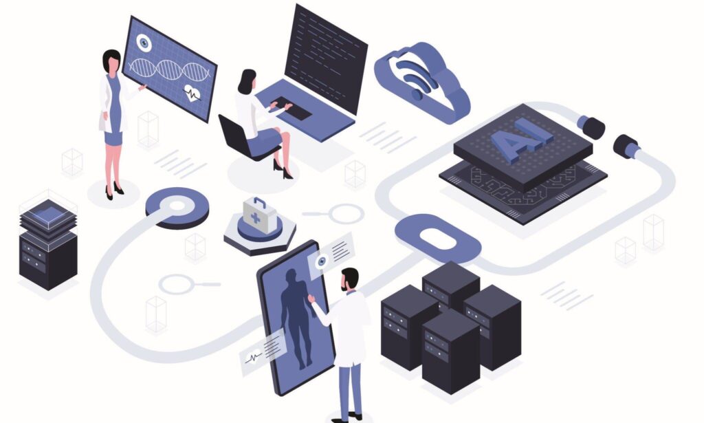
What is the goal, target audience, and areas of digital health it addresses?
The main goal of this research is to validate a fusion algorithm that integrates Magnetic Resonance Imaging (MRI) and 3D scan to improve tumor localization in breast cancer patients. The target audience includes surgeons, radiologists, and clinical researchers. The study addresses the areas of medical imaging, image processing, computer-assisted surgery, and augmented reality for surgery.
What is the context?
Breast-conserving surgery aims to remove tumors while preserving as much healthy breast tissue as possible, ensuring optimal aesthetic outcomes that are critical for a patient’s quality of life. To achieve this objective, precise location of the tumor is necessary during surgery, which is normally performed with the patient lying face-up (supine position).
To plan the procedure, surgeons rely on medical imaging technologies that often provide limited perspectives due to the conditions under which the images are captured. For instance, MRI scans are performed with the patient lying face down (prone position), causing the breast to compress and deform, while 3D scans capture the natural shape of the breast in the upright position. Since neither imaging method reflects the breast’s actual position during surgery, merging internal radiological images (e.g., MRI) with external surface images (e.g., 3D scans) into a unified model is essential to create a realistic representation of the breast. However, aligning these datasets is challenging not only because the images are captured in different positions but also due to the limited availability of anatomical reference points for alignment. To solve this issue, artificial reference points, such as marks made with black markers, can be used; however, these points are not visible on MRI, as it only captures internal structures. Alternatively, cod liver oil pills offer a low-cost and readily available solution, providing contrast on MRI due to their fat content.
What are the current approaches?
Current approaches to breast image fusion typically focus on aligning radiological or surface imaging data independently, rather than integrating both into a unified 3D model.
One approach used is the biomechanical models, which simulate how breast tissue moves and changes shape between different positions. These models are based on mathematical equations to predict tissue behavior in detail, generating realistic simulations. However, they are computationally intensive, prone to introducing distortions in breast shape, and often unable to reflect the unique properties of each patient’s breast.
Another method involves non-physical models like Free Form Deformation (FFD), which use a flexible “grid” of control points to adjust the shape of images. This technique directly bends and stretches images to align them, offering a faster and simpler alternative to biomechanical models. However, FFD has limitations in terms of physical realism and may generate less accurate results as it does not consider the natural behavior of breast tissue.
Combining these two methods—using biomechanical models for more realistic simulations and FFD for alignment adjustments—shows promise, but still faces challenges, such as limited clinical validation.
What does innovation consist of? How is the impact of this study assessed?
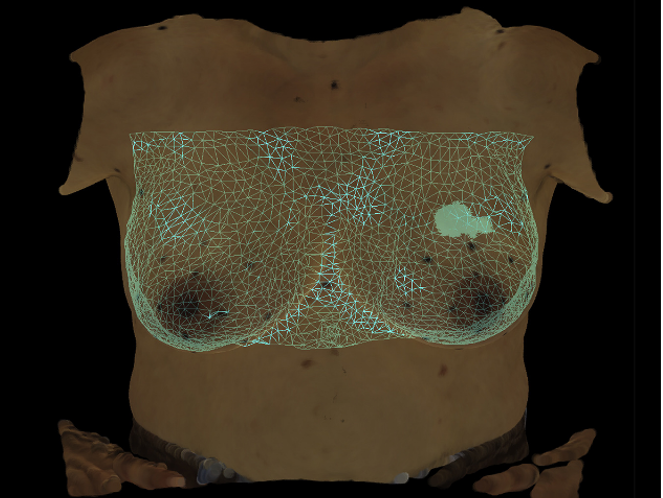
The innovation in this study lies in the development and validation of a fusion algorithm that integrates MRI data and 3D scan data to create unified, patient-specific 3D breast models, intended to support surgical planning.
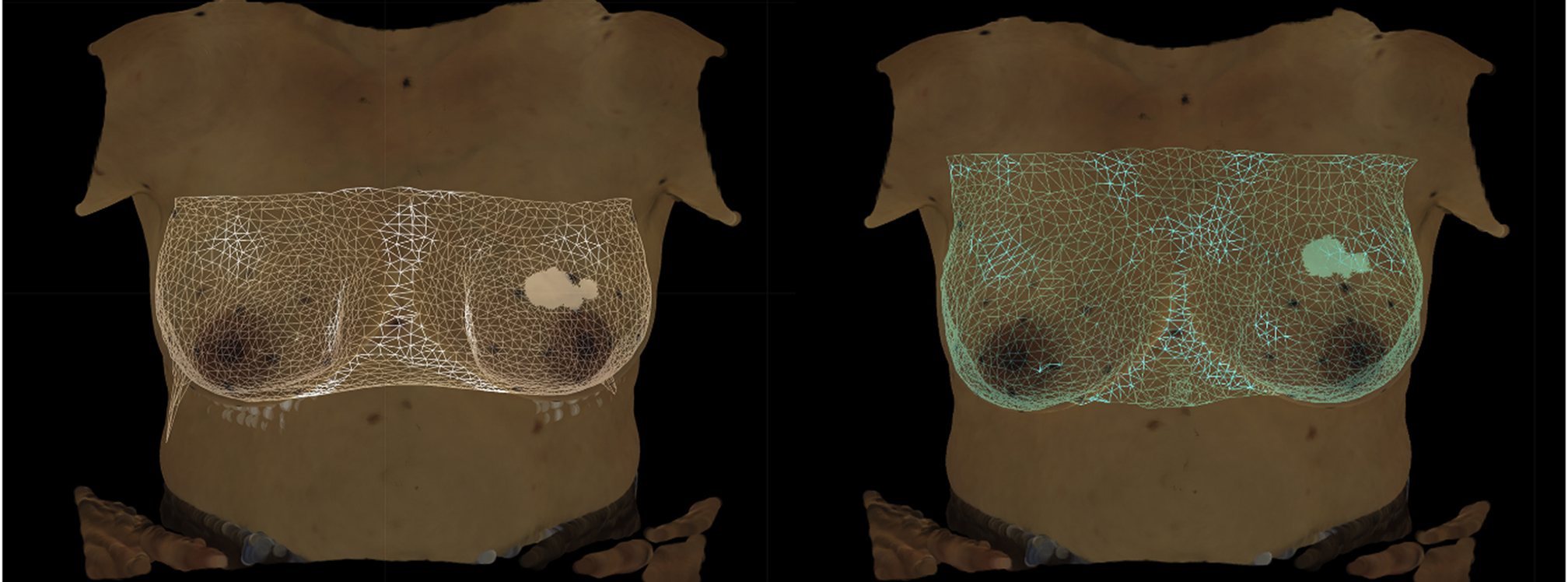
The protocol begins with data acquisition, where both MRI scans and 3D scans are collected. Then, radiologists use the Horos R software to identify and outline tumors in the MRI scans, calculating their volume to create a 3D tumor model that is, then, overlaid onto the original MRI dataset. To prepare for fusion, two MRI datasets are created with the tumor marked: MRIprone (an image with the original MRI without biomechanical processing) and MRIup (an image adjusted with a biomechanical model to simulate the upright position obtained in the 3D scan).
Both MRIprone and MRIup datasets are submitted to the fusion algorithm, which integrates them with the 3D scans using two types of alignment strategies: single breast fusion, where each breast is fused independently with the corresponding side of the breast in the 3D scan, and full torso fusion, where both breasts and the torso are fused as a single unit. The alignment performed involves superimposing the contours of both sets of images. This step relies on anatomical reference points as well as artificial reference points, known as Breast Surface Markers (BSM). These points are created using either black permanent markers on the breast and torso before 3D scan acquisition or cod liver oil pills placed in the same positions before MRI acquisition, ensuring correspondence between the two imaging modalities.
Then, the fusion algorithm applies FFD adjustments to compensate for residual differences in breast shape caused by factors such as variations in imaging positions, soft tissue deformations, and gravitational effects. During FFD, the tumor location, initially outlined in the MRI data, is carefully adjusted to align with the fused 3D breast model.
Seven breast cancer patients eligible for breast-conserving surgery at the Champalimaud Clinical Center in Portugal participated in the validation of the fusion algorithm with the application of BSM. The aim was also to identify the most effective approach for tumor detection by the algorithm, comparing the fusion of 3D scans with data obtained in MRIprone and MRIup, as well as individual breast fusion with full torso fusion alignment.
The performance of the technology was evaluated using two complementary methods: Target Registration Error (TRE) and qualitative tumor localization validation. TRE quantifies the alignment accuracy between the MRI and 3D scan by measuring the Euclidean distance—a straight-line distance in 3D space—between corresponding BSM identified in both datasets, providing an objective metric for fusion precision. Additionally, qualitative validation was conducted by a breast surgeon, who compared the tumor’s location in the fused 3D breast model against clinical records, surgical annotations, and visible skin markers to ensure the tumor’s position was accurately represented in the final model.
What are the main results? What is the impact of these results? What is the future of this technology?
The fusion algorithm successfully created accurate 3D breast models and demonstrated robust performance across different patient anatomies, as it was not significantly influenced by variations in breast volume.
The results showed that single breast fusion with MRIprone provided more precise tumor localization despite having slightly higher TRE values, indicating a small mismatch in aligning the datasets. Specifically, the TRE for single breast fusion with MRIprone was 26.26 ± 6.61 mm, compared to a lower TRE of 18.5 ± 3.88 mm for single breast fusion with MRIup. However, the use of a biomechanical model in MRIup introduced artifacts such as axial elongation and lateral displacement, which negatively impacted tumor positioning accuracy. Qualitative analysis further highlighted that tumor localization was more accurate in 80% of cases with MRIprone. In contrast, the full torso fusion approaches exhibited the poorest overall performance.
The results pave the way for more patient-specific breast cancer imaging in clinical practice. The validated algorithm eliminates the need for complex biomechanical models, offering a simpler solution for tumor localization. This approach supports automation, improves preoperative planning, and enhances surgical precision in breast-conserving treatments. The ability to generate precise 3D models also enables the integration of augmented reality, providing surgeons with real-time visualization of tumor location during operations, which should improve patient outcomes.
The future involves obtaining MRI images in the supine position, bringing imaging acquisition conditions closer to surgical scenarios, which will allow for improved tumor localization accuracy. It is also necessary to expand datasets with more participants and integrate machine learning to enhance algorithm performance and enable the prediction of breast shape transformations. The use of augmented reality glasses, with the overlay of 3D breast models in real-world contexts, could revolutionize surgery by allowing direct visualization of tumor locations, making procedures more precise and efficient.
Would you like to know all the details?

Autonomous Robotics System for Autism Therapy
Autism spectrum disorder is a neurodevelopmental condition with significant clinical, social and economic repercussions throughout life. According to the World Health Organization, it is estimated…
Virtual Reality Exergames as a Tool for Diagnosing Eye Diseases
Eye diseases represent a growing public health challenge in Portugal, significantly compromising the population’s quality of life. The increase in their prevalence is associated with…

Improving Efficiency in the Clinical Follow-up for Covid-19 Cases With a Digital Platform
COVID-19, caused by the SARS-CoV-2 virus, is a highly contagious disease with the potential to cause serious complications, requiring the isolation of infected individuals and…

The Rising Threat of Antibiotic-resistant Klebsiella in Portuguese Hospitals
Healthcare-associated infections pose a serious public health threat, as they are acquired during medical treatments or hospital stays, often leading to prolonged hospitalizations, high costs…

Deep Neural Networks And The Future Of Early Detection Of Alzheimer’s Disease
Alzheimer’s disease is the most common form of dementia, affecting more than 55 million people globally and accounting for around 70 percent of dementia cases….

Mobile Application to Improve Workflows in Nursing Homes
Portugal has one of the highest aging populations in the world, placing increasing pressure on elderly care services, especially in nursing homes. Healthcare professionals in…

The Challenges of Data Protection in Digital Health Platforms for the Elderly
Demographic ageing poses significant challenges to healthcare systems, intensifying the pressure on infrastructures and human resources. It is estimated that by 2050 the elderly population…
Facilitating Epilepsy Diagnosis With a Wireless and Wearable EEG System
Paroxysmal diseases are characterized by sudden, episodic conditions that cause temporary changes in the body. Among them, epilepsy stands out for causing synchronous and uncontrolled…
Automatic Segmentation of Blood Vessels in Carotid Ultrasound Images
Vascular diseases, such as carotid stenosis (narrowing of the carotid arteries, which connect the heart to the brain, caused by the accumulation of fatty atheroma…

Impact of Robotherapy-PARO on Elderly People With Dementia in Portugal
Aging is a gradual, multifactorial and continuous process characterized by the progressive loss of biological function and degeneration associated with the onset of age-related diseases….
New Era of Interoperability in Healthcare Systems
The growing use of electronic health records, digital diagnostic systems and remote monitoring technologies has led to a significant increase in the volume and complexity…
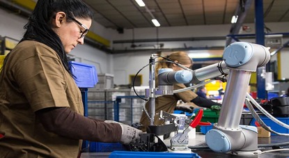
Collaborative Robotics Improves Working Conditions
Workers face growing challenges in the industrial environment. Among the most critical are fatigue and inappropriate postures, often associated with repetitive tasks and working conditions…
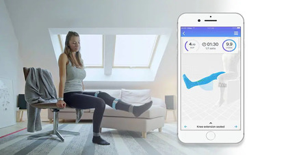
The Role of Mobile Technologies in the Monitoring and Rehabilitation of Peripheral Arterial Disease
PAD is a prevalent chronic condition, affecting approximately 200 million individuals globally, characterized by obstruction of the peripheral arteries, especially in the lower extremities, due…
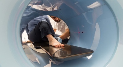
Incorporation of Digital Implants Into CT Images to Plan Orthopedic Surgery
Orthopedic surgery addresses conditions of the musculoskeletal system to alleviate pain, restore function, and enhance the patient’s quality of life. Its success relies on meticulous…

Digital Health at the Top of the National Poliempreende 2024 Results
Poliempreende is a consolidated national network for encouraging entrepreneurship in higher education in Portugal, with two decades of existence. Focused on promoting innovation, the competition…
Digital Solution Facilitates Interaction Between Users and Health Professionals
Many patients face difficulties scheduling medical appointments in hospital units, and, when successful, they often endure long waiting times to be attended. This situation is…
The Impact of Calm Computing Integration on the Clinical Process
In recent years, digital transformation in healthcare has played a crucial role, driven by the exponential increase in medical data. This ranges from administrative information…
ULS Almada-Seixal Revolutionizes With the Region’s First Surgical Robot
In recent years, ULSAS has been gradually implementing robotic systems, reinforcing its commitment to innovation and improving healthcare. Recently, the institution acquired a state-of-the-art robotic…

Online Intervention Aims to Prevent Anxiety in the General Population
Anxiety disorders are a global problem, affecting 300 million people worldwide and placing significant pressure on individuals and healthcare systems. In Europe alone, the economic…
Rehabilitation of Facial Paralysis Through Virtual Assistants
Facial paralysis, defined by the inability to move one or both sides of the face, has an incidence of 20 to 30 cases per 100,000…
Detection of Anxiety and Panic Attacks in Real Time
The growing number of people with anxiety disorders, along with increased awareness of mental health, drives the need for new technological tools that provide remote…
A Novel Approach for Robotic-assisted Tele-echography
Currently, robotic systems for ultrasound diagnostic procedures fall into two main categories: portable robots that require manual positioning and fully autonomous robotic systems that independently…

From Big Data to Big Decisions: How AI Stratifies Cancer Cases by Risk Factors
The CLARIFY Decision Support Platform (DSP) is a responsive web application designed to support decision-making in cancer care through real-time data integration and predictive analytics….

From “Free Text” to Structured Clinical Data: the Foundation for Clinical Decision Support Systems
Currently, the practice of recording clinical information in “free text” offers flexibility, but hinders automatic data extraction, limiting the application of analytical models. Most records…
Artificial Intelligence used in Depression Detection in Cancer Survivors
The goal of the FAITH project (Federated Artificial Intelligence solution for moniToring mental Health status after cancer treatment) is to remotely identify and predict depressive…

Integration of SONHO v2 and SClínico Systems at ULS of Coimbra to Improve Healthcare Services
With more than half a million hospital medical consultations carried out in the first half of 2024, the ULS of Coimbra stands out as an…

Elderly Care Ecosystem: an Innovative Platform for Personalized and Efficient Services
The Elderly Care Ecosystem (ECE) is an integration of various digital health technologies, exploring the areas of telehealth and predictive analytics. The goal of this…

Innovative technology that subconsciously relieves anxiety through a scarf
The SCAARF technology aims to offer an alternative method to alleviate anxiety symptoms in a non-intrusive and subconscious way. This technology is an innovative idea…

Digital Health Interventions: Equity in Hypertension Care for Everyone
Nearly half of all adults in the United States have hypertension, one of the leading risk factors for cardiovascular disease, and only about a quarter…
Personalization and Technology in Diabetes Management
IPDM has significant potential to improve diabetes management and drive health system reforms to become high-performing, effective, equitable, accessible, and sustainable. Evidence and good practices…
Negotiations on the European Health Data Space Advance With the Participation of the SPMS
The European Health Data Space will be a common health data sharing system across the European Union. It foresees the use of data for purposes…
Secretary of State Margarida Tavares Emphasizes Digital Innovation in Health Promotion
Margarida Tavares spoke at the opening of the conference ” O Digital na promoção contínua da saúde e do bem-estar”, organized by the Associação para…
ARS Algarve Modernizes Radiology With AI and New Data Center
The radiology service of ARS Algarve has already performed nearly 29,000 exams using Artificial Intelligence (AI) technology. In recent years, there has been a significant…

European Health Data Space: Unified Access To Health Data In The EU
The COVID-19 pandemic highlighted the importance of digital services in health, but complex rules and increasing cyberattacks make it difficult to share data across Member…

European Commission Amends Digital Europe Programme With an Investment of €762.7 Million
The European Commission has amended the Digital Europe Programme work programmes 2023-2024, investing an additional €762.7 million in Europe’s digital transition and cybersecurity. The digital…
SPMS Integrates the TEF-Health Initiative
SPMS participates in the TEF-Health initiative as a partner in a consortium composed of 51 entities from 9 European Union countries. This action is co-financed…

FMUP Creates Inhealth Junior Academy for High School Students
The InHealth Junior Academy — Academia Júnior de Inovação em Saúde is an initiative of the Departamento de Medicina da Comunidade, Informação e Decisão em…

SPMS Represents Portugal as Vice-president of GDHP
The GDHP is an intergovernmental organization in the digital health sector that facilitates cooperation and collaboration between government representatives and the World Health Organization (WHO)….
Digital Transformation of Health at INCoDe.2030 in Tomar
The “National Digital Skills Initiative e.2030, Portugal” (INCoDe.2030) is an initiative that aims to improve the Portuguese population’s level of digital skills, placing Portugal at…
Braga Hospital Evaluates Memory With Interactive Game in Patients With Multiple Sclerosis
Multiple Sclerosis is known as a chronic disease of the central nervous system, with a wide variety of motor and sensory symptoms that can lead…

Neurosurgery Teleconsultation Wins Innovation Award
The aim of the BI Award for Innovation in Healthcare is to recognize innovative projects in the healthcare sector that improve the quality of life…
HealthData@PT: New SPMS Initiative for Health Data
Action HealthData@PT is launched in the context of the implementation of the European Health Data Space, and is an initiative approved by the European Commission…
Do you have an innovative idea in healthcare field?
Share it with us and see it come to life.
We will help bring your projects to life!


Home / Publications / Publication
IMPROVEMENT IN BREAST TUMOR LOCALIZATION WITH AN IMAGE FUSION ALGORITHM
Publication Type: Article Summary
Original title: 3D digital breast cancer models with multimodal fusion algorithms
Article publication date: February 2020
Source: Repositório da Universidade Nova de Lisboa
Authors: Sílvia Bessa, Pedro Gouveia, Pedro Carvalho, Cátia Rodrigues, Nuno Silva, Fátima Cardoso, Jaime Cardoso, Hélder Oliveira & Maria João Cardoso
What is the goal, target audience, and areas of digital health it addresses?
The main goal of this research is to validate a fusion algorithm that integrates Magnetic Resonance Imaging (MRI) and 3D scan to improve tumor localization in breast cancer patients. The target audience includes surgeons, radiologists, and clinical researchers. The study addresses the areas of medical imaging, image processing, computer-assisted surgery, and augmented reality for surgery.
What is the context?
Breast-conserving surgery aims to remove tumors while preserving as much healthy breast tissue as possible, ensuring optimal aesthetic outcomes that are critical for a patient’s quality of life. To achieve this objective, precise location of the tumor is necessary during surgery, which is normally performed with the patient lying face-up (supine position).
To plan the procedure, surgeons rely on medical imaging technologies that often provide limited perspectives due to the conditions under which the images are captured. For instance, MRI scans are performed with the patient lying face down (prone position), causing the breast to compress and deform, while 3D scans capture the natural shape of the breast in the upright position. Since neither imaging method reflects the breast’s actual position during surgery, merging internal radiological images (e.g., MRI) with external surface images (e.g., 3D scans) into a unified model is essential to create a realistic representation of the breast. However, aligning these datasets is challenging not only because the images are captured in different positions but also due to the limited availability of anatomical reference points for alignment. To solve this issue, artificial reference points, such as marks made with black markers, can be used; however, these points are not visible on MRI, as it only captures internal structures. Alternatively, cod liver oil pills offer a low-cost and readily available solution, providing contrast on MRI due to their fat content.
What are the current approaches?
Current approaches to breast image fusion typically focus on aligning radiological or surface imaging data independently, rather than integrating both into a unified 3D model.
One approach used is the biomechanical models, which simulate how breast tissue moves and changes shape between different positions. These models are based on mathematical equations to predict tissue behavior in detail, generating realistic simulations. However, they are computationally intensive, prone to introducing distortions in breast shape, and often unable to reflect the unique properties of each patient’s breast.
Another method involves non-physical models like Free Form Deformation (FFD), which use a flexible “grid” of control points to adjust the shape of images. This technique directly bends and stretches images to align them, offering a faster and simpler alternative to biomechanical models. However, FFD has limitations in terms of physical realism and may generate less accurate results as it does not consider the natural behavior of breast tissue.
Combining these two methods—using biomechanical models for more realistic simulations and FFD for alignment adjustments—shows promise, but still faces challenges, such as limited clinical validation.
What does innovation consist of? How is the impact of this study assessed?
The innovation in this study lies in the development and validation of a fusion algorithm that integrates MRI data and 3D scan data to create unified, patient-specific 3D breast models, intended to support surgical planning.
The protocol begins with data acquisition, where both MRI scans and 3D scans are collected. Then, radiologists use the Horos R software to identify and outline tumors in the MRI scans, calculating their volume to create a 3D tumor model that is, then, overlaid onto the original MRI dataset. To prepare for fusion, two MRI datasets are created with the tumor marked: MRIprone (an image with the original MRI without biomechanical processing) and MRIup (an image adjusted with a biomechanical model to simulate the upright position obtained in the 3D scan).
Both MRIprone and MRIup datasets are submitted to the fusion algorithm, which integrates them with the 3D scans using two types of alignment strategies: single breast fusion, where each breast is fused independently with the corresponding side of the breast in the 3D scan, and full torso fusion, where both breasts and the torso are fused as a single unit. The alignment performed involves superimposing the contours of both sets of images. This step relies on anatomical reference points as well as artificial reference points, known as Breast Surface Markers (BSM). These points are created using either black permanent markers on the breast and torso before 3D scan acquisition or cod liver oil pills placed in the same positions before MRI acquisition, ensuring correspondence between the two imaging modalities.
Then, the fusion algorithm applies FFD adjustments to compensate for residual differences in breast shape caused by factors such as variations in imaging positions, soft tissue deformations, and gravitational effects. During FFD, the tumor location, initially outlined in the MRI data, is carefully adjusted to align with the fused 3D breast model.
Seven breast cancer patients eligible for breast-conserving surgery at the Champalimaud Clinical Center in Portugal participated in the validation of the fusion algorithm with the application of BSM. The aim was also to identify the most effective approach for tumor detection by the algorithm, comparing the fusion of 3D scans with data obtained in MRIprone and MRIup, as well as individual breast fusion with full torso fusion alignment.
The performance of the technology was evaluated using two complementary methods: Target Registration Error (TRE) and qualitative tumor localization validation. TRE quantifies the alignment accuracy between the MRI and 3D scan by measuring the Euclidean distance—a straight-line distance in 3D space—between corresponding BSM identified in both datasets, providing an objective metric for fusion precision. Additionally, qualitative validation was conducted by a breast surgeon, who compared the tumor’s location in the fused 3D breast model against clinical records, surgical annotations, and visible skin markers to ensure the tumor’s position was accurately represented in the final model.
What are the main results? What is the impact of these results? What is the future of this technology?
The fusion algorithm successfully created accurate 3D breast models and demonstrated robust performance across different patient anatomies, as it was not significantly influenced by variations in breast volume.
The results showed that single breast fusion with MRIprone provided more precise tumor localization despite having slightly higher TRE values, indicating a small mismatch in aligning the datasets. Specifically, the TRE for single breast fusion with MRIprone was 26.26 ± 6.61 mm, compared to a lower TRE of 18.5 ± 3.88 mm for single breast fusion with MRIup. However, the use of a biomechanical model in MRIup introduced artifacts such as axial elongation and lateral displacement, which negatively impacted tumor positioning accuracy. Qualitative analysis further highlighted that tumor localization was more accurate in 80% of cases with MRIprone. In contrast, the full torso fusion approaches exhibited the poorest overall performance.
The results pave the way for more patient-specific breast cancer imaging in clinical practice. The validated algorithm eliminates the need for complex biomechanical models, offering a simpler solution for tumor localization. This approach supports automation, improves preoperative planning, and enhances surgical precision in breast-conserving treatments. The ability to generate precise 3D models also enables the integration of augmented reality, providing surgeons with real-time visualization of tumor location during operations, which should improve patient outcomes.
The future involves obtaining MRI images in the supine position, bringing imaging acquisition conditions closer to surgical scenarios, which will allow for improved tumor localization accuracy. It is also necessary to expand datasets with more participants and integrate machine learning to enhance algorithm performance and enable the prediction of breast shape transformations. The use of augmented reality glasses, with the overlay of 3D breast models in real-world contexts, could revolutionize surgery by allowing direct visualization of tumor locations, making procedures more precise and efficient.
Would you like to know all the details?
Autonomous Robotics System for Autism Therapy
Autism spectrum disorder is a neurodevelopmental condition with significant clinical, social and economic repercussions throughout life. According to the World Health Organization, it is estimated to affect approximately 1 in 160 children worldwide. Its origin…
Virtual Reality Exergames as a Tool for Diagnosing Eye Diseases
Eye diseases represent a growing public health challenge in Portugal, significantly compromising the population’s quality of life. The increase in their prevalence is associated with various factors, such as demographic ageing, excessive use of digital…
Improving Efficiency in the Clinical Follow-up for Covid-19 Cases With a Digital Platform
COVID-19, caused by the SARS-CoV-2 virus, is a highly contagious disease with the potential to cause serious complications, requiring the isolation of infected individuals and appropriate clinical follow-up. While severe cases require hospitalization, patients with…
The Rising Threat of Antibiotic-resistant Klebsiella in Portuguese Hospitals
Healthcare-associated infections pose a serious public health threat, as they are acquired during medical treatments or hospital stays, often leading to prolonged hospitalizations, high costs for healthcare systems and high mortality rates. Portugal has one…
Deep Neural Networks And The Future Of Early Detection Of Alzheimer’s Disease
Alzheimer’s disease is the most common form of dementia, affecting more than 55 million people globally and accounting for around 70 percent of dementia cases. In Portugal, it is estimated that 200,000 people live with…
Mobile Application to Improve Workflows in Nursing Homes
Portugal has one of the highest aging populations in the world, placing increasing pressure on elderly care services, especially in nursing homes. Healthcare professionals in these facilities are often overwhelmed due to the increasing number…
The Challenges of Data Protection in Digital Health Platforms for the Elderly
Demographic ageing poses significant challenges to healthcare systems, intensifying the pressure on infrastructures and human resources. It is estimated that by 2050 the elderly population will exceed 2 billion people, making it imperative to implement…
Facilitating Epilepsy Diagnosis With a Wireless and Wearable EEG System
Paroxysmal diseases are characterized by sudden, episodic conditions that cause temporary changes in the body. Among them, epilepsy stands out for causing synchronous and uncontrolled neuronal discharges, resulting in recurrent and unprovoked seizures. These seizures…
Automatic Segmentation of Blood Vessels in Carotid Ultrasound Images
Vascular diseases, such as carotid stenosis (narrowing of the carotid arteries, which connect the heart to the brain, caused by the accumulation of fatty atheroma plaques), cerebrovascular accidents (CVA) (sudden interruption of blood flow to…
Impact of Robotherapy-PARO on Elderly People With Dementia in Portugal
Aging is a gradual, multifactorial and continuous process characterized by the progressive loss of biological function and degeneration associated with the onset of age-related diseases. In Portugal, the aging of the population is particularly noticeable,…
New Era of Interoperability in Healthcare Systems
The growing use of electronic health records, digital diagnostic systems and remote monitoring technologies has led to a significant increase in the volume and complexity of health data. This increase intensifies the need for continuous,…
Collaborative Robotics Improves Working Conditions
Workers face growing challenges in the industrial environment. Among the most critical are fatigue and inappropriate postures, often associated with repetitive tasks and working conditions that lack ergonomic suitability. These factors represent significant risks for…
The Role of Mobile Technologies in the Monitoring and Rehabilitation of Peripheral Arterial Disease
PAD is a prevalent chronic condition, affecting approximately 200 million individuals globally, characterized by obstruction of the peripheral arteries, especially in the lower extremities, due to the formation of atherosclerotic plaques, which compromise blood flow…
Incorporation of Digital Implants Into CT Images to Plan Orthopedic Surgery
Orthopedic surgery addresses conditions of the musculoskeletal system to alleviate pain, restore function, and enhance the patient’s quality of life. Its success relies on meticulous pre-operative planning that incorporates clinical data and patient-specific imaging to…
Digital Health at the Top of the National Poliempreende 2024 Results
Poliempreende is a consolidated national network for encouraging entrepreneurship in higher education in Portugal, with two decades of existence. Focused on promoting innovation, the competition has had a significant impact on the national economy, with…
Digital Solution Facilitates Interaction Between Users and Health Professionals
Many patients face difficulties scheduling medical appointments in hospital units, and, when successful, they often endure long waiting times to be attended. This situation is aggravated by problems such as the incompatibility of schedules between…
The Impact of Calm Computing Integration on the Clinical Process
In recent years, digital transformation in healthcare has played a crucial role, driven by the exponential increase in medical data. This ranges from administrative information to detailed records of diagnoses, laboratory tests, medical images and…
ULS Almada-Seixal Revolutionizes With the Region’s First Surgical Robot
In recent years, ULSAS has been gradually implementing robotic systems, reinforcing its commitment to innovation and improving healthcare. Recently, the institution acquired a state-of-the-art robotic system, developed under the concept of an ‘immersive intuitive interface’,…
Online Intervention Aims to Prevent Anxiety in the General Population
Anxiety disorders are a global problem, affecting 300 million people worldwide and placing significant pressure on individuals and healthcare systems. In Europe alone, the economic impact reached 74.380 million in 2010, with 62.2% attributed to…
Rehabilitation of Facial Paralysis Through Virtual Assistants
Facial paralysis, defined by the inability to move one or both sides of the face, has an incidence of 20 to 30 cases per 100,000 people annually. This condition often causes facial weakness, difficulties in…
Detection of Anxiety and Panic Attacks in Real Time
The growing number of people with anxiety disorders, along with increased awareness of mental health, drives the need for new technological tools that provide remote and continuous monitoring of anxiety and panic disorders. Thus, the…
A Novel Approach for Robotic-assisted Tele-echography
Currently, robotic systems for ultrasound diagnostic procedures fall into two main categories: portable robots that require manual positioning and fully autonomous robotic systems that independently control the ultrasound probe’s orientation and positioning. Portable robots rely…
From Big Data to Big Decisions: How AI Stratifies Cancer Cases by Risk Factors
The CLARIFY Decision Support Platform (DSP) is a responsive web application designed to support decision-making in cancer care through real-time data integration and predictive analytics. Built on Big Data Europe, the platform integrates a variety…
From “Free Text” to Structured Clinical Data: the Foundation for Clinical Decision Support Systems
Currently, the practice of recording clinical information in “free text” offers flexibility, but hinders automatic data extraction, limiting the application of analytical models. Most records are unstructured, and the use of non-standard abbreviations increases ambiguity,…
Artificial Intelligence used in Depression Detection in Cancer Survivors
The goal of the FAITH project (Federated Artificial Intelligence solution for moniToring mental Health status after cancer treatment) is to remotely identify and predict depressive symptoms in cancer survivors using a federated machine learning approach…
Integration of SONHO v2 and SClínico Systems at ULS of Coimbra to Improve Healthcare Services
With more than half a million hospital medical consultations carried out in the first half of 2024, the ULS of Coimbra stands out as an institution dedicated to integrated, high-quality and patient-centered healthcare, with 8…
Elderly Care Ecosystem: an Innovative Platform for Personalized and Efficient Services
The Elderly Care Ecosystem (ECE) is an integration of various digital health technologies, exploring the areas of telehealth and predictive analytics. The goal of this ecosystem is to improve the quality of life for elderly…
Innovative technology that subconsciously relieves anxiety through a scarf
The SCAARF technology aims to offer an alternative method to alleviate anxiety symptoms in a non-intrusive and subconscious way. This technology is an innovative idea in the field of digital health and wearable technology, designed…
Digital Health Interventions: Equity in Hypertension Care for Everyone
Nearly half of all adults in the United States have hypertension, one of the leading risk factors for cardiovascular disease, and only about a quarter (24%) of those people have their hypertension under control. Studies…
Personalization and Technology in Diabetes Management
IPDM has significant potential to improve diabetes management and drive health system reforms to become high-performing, effective, equitable, accessible, and sustainable. Evidence and good practices inspire health system transformation. Adopting person-centred approaches like co-creation and…
Negotiations on the European Health Data Space Advance With the Participation of the SPMS
The European Health Data Space will be a common health data sharing system across the European Union. It foresees the use of data for purposes that benefit people and society. It will ensure citizens have…
Secretary of State Margarida Tavares Emphasizes Digital Innovation in Health Promotion
Margarida Tavares spoke at the opening of the conference ” O Digital na promoção contínua da saúde e do bem-estar”, organized by the Associação para a Promoção e Desenvolvimento da Sociedade da Informação (APDSI) and…
ARS Algarve Modernizes Radiology With AI and New Data Center
The radiology service of ARS Algarve has already performed nearly 29,000 exams using Artificial Intelligence (AI) technology. In recent years, there has been a significant investment in image digitization and data storage, as well as…
European Health Data Space: Unified Access To Health Data In The EU
The COVID-19 pandemic highlighted the importance of digital services in health, but complex rules and increasing cyberattacks make it difficult to share data across Member States; the EHDS, based on several regulations, provides tailor-made rules…
European Commission Amends Digital Europe Programme With an Investment of €762.7 Million
The European Commission has amended the Digital Europe Programme work programmes 2023-2024, investing an additional €762.7 million in Europe’s digital transition and cybersecurity. The digital transition is the main work programme with a budget of…
SPMS Integrates the TEF-Health Initiative
SPMS participates in the TEF-Health initiative as a partner in a consortium composed of 51 entities from 9 European Union countries. This action is co-financed by the European Commission and has a duration of five…
FMUP Creates Inhealth Junior Academy for High School Students
The InHealth Junior Academy — Academia Júnior de Inovação em Saúde is an initiative of the Departamento de Medicina da Comunidade, Informação e Decisão em Saúde da Faculdade de Medicina da Universidade do Porto (FMUP)….
SPMS Represents Portugal as Vice-president of GDHP
The GDHP is an intergovernmental organization in the digital health sector that facilitates cooperation and collaboration between government representatives and the World Health Organization (WHO). Its purpose is to foster policymaking that promote the digitalization…
Digital Transformation of Health at INCoDe.2030 in Tomar
The “National Digital Skills Initiative e.2030, Portugal” (INCoDe.2030) is an initiative that aims to improve the Portuguese population’s level of digital skills, placing Portugal at the level of the most advanced European countries in this…
Braga Hospital Evaluates Memory With Interactive Game in Patients With Multiple Sclerosis
Multiple Sclerosis is known as a chronic disease of the central nervous system, with a wide variety of motor and sensory symptoms that can lead to work disability, socioeconomic burden, and reduced quality of life…
Neurosurgery Teleconsultation Wins Innovation Award
The aim of the BI Award for Innovation in Healthcare is to recognize innovative projects in the healthcare sector that improve the quality of life for the Portuguese people. In 2021, the specific theme was…
HealthData@PT: New SPMS Initiative for Health Data
Action HealthData@PT is launched in the context of the implementation of the European Health Data Space, and is an initiative approved by the European Commission under the EU4Health 2021-2027 programme. This initiative contributes to the…
Do you have an innovative idea in healthcare field?
Share it with us and see it come to life.
We will help bring your projects to life!