
Vascular diseases, such as carotid stenosis (narrowing of the carotid arteries, which connect the heart to the brain, caused by the accumulation of fatty atheroma plaques), cerebrovascular accidents (CVA) (sudden interruption of blood flow to the brain), atherosclerosis (accumulation of atheroma plaques in the arteries, leading to arterial hardening and narrowing), deep vein thrombosis (formation of blood clots in deep veins), and cervical arterial dissections (tears in the inner layer of the neck arteries), represent a significant challenge to global public health due to their high prevalence and clinical impact. Vessels
Home / Publications / Publication
Home / Publications / Publication
AUTOMATIC SEGMENTATION OF BLOOD VESSELS IN CAROTID ULTRASOUND IMAGES
Publication type: Article Summary
Original title: Vessel Detection In Carotid Ultrasound Images Using Artificial Neural Networks
Article publication date: July 2018
Source: Proceedings of the 6th International Conference Integrity, Reliability and Failure
Authors: Catarina F. Castro, Carlos C. António & Luísa C. Sousa
What is the goal, target audience, and areas of digital health it addresses?
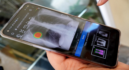
The study aims to automatically identify blood vessels and segment carotid ultrasound images, improving diagnostic accuracy and clinical efficiency. The target audience includes healthcare professionals, such as radiologists and angiologists, as well as healthcare institutions and researchers in the fields of Artificial Intelligence (AI) and medical image processing. The study falls within the areas of digital health, with a focus on the application of AI and diagnostic support technologies.
What is the context?
Vascular diseases, such as carotid stenosis (narrowing of the carotid arteries, which connect the heart to the brain, caused by the accumulation of fatty atheroma plaques), cerebrovascular accidents (CVA) (sudden interruption of blood flow to the brain), atherosclerosis (accumulation of atheroma plaques in the arteries, leading to arterial hardening and narrowing), deep vein thrombosis (formation of blood clots in deep veins), and cervical arterial dissections (tears in the inner layer of the neck arteries), represent a significant challenge to global public health due to their high prevalence and clinical impact. Carotid stenosis is a major risk factor for CVA, affecting millions of people annually. Atherosclerosis, the leading cause of cardiovascular complications, is increasing with population ageing and risk factors such as obesity, sedentary lifestyles, and hypertension. Deep vein thrombosis, which occurs in 1 to 2 individuals per 1000 annually, can lead to pulmonary embolism (blockage of a pulmonary artery by an embolus), a severe and often fatal complication. Although less common, cervical arterial dissections are a significant cause of CVA in young adults and are frequently underdiagnosed until severe complications arise.
These conditions highlight the urgent need for advanced diagnostic tools that enable early detection and accurate assessment of these pathologies, contributing to improved clinical management and reduced complications.
What are the current approaches?
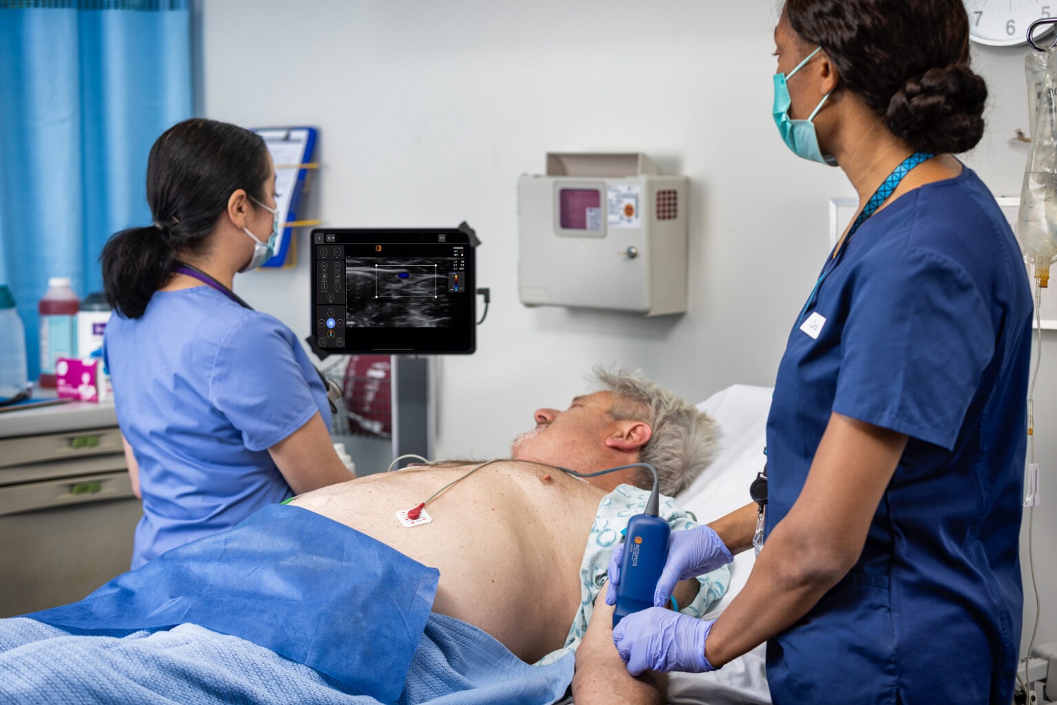
Current approaches for the diagnosis and monitoring of vascular diseases integrate advanced imaging techniques, computational methods, and clinical tools to enhance accuracy and efficiency in diagnostic outcomes. Doppler ultrasound is widely used due to its non-invasive nature, speed, and low cost, proving effective in assessing blood flow and detecting arterial obstructions. More sophisticated methods, such as computed tomography angiography and magnetic resonance angiography, provide high-resolution imaging that facilitates the detection of atheroma plaques, arterial dissections, and vascular occlusions (blockages in blood vessels). Additionally, transcranial Doppler ultrasound plays a crucial role in real-time monitoring of cerebral blood flow, proving useful in assessing conditions such as intracranial stenosis (narrowing of arteries within the skull) and other cerebrovascular disorders.
Despite their clinical utility, these techniques present limitations, including operator-dependent settings, image irregularities, and challenges in distinguishing vascular from non-vascular structures in transverse images. These factors can compromise diagnostic accuracy, particularly in cases with image artifacts or noise. Such limitations highlight the urgent need for automated, more robust, and precise solutions that can minimize variability and errors associated with manual analysis, ensuring greater consistency and accuracy in the diagnosis and monitoring of vascular diseases.
What does innovation consist of?
The innovation of this study lies in the development of an advanced methodology for the automatic segmentation of blood vessels in transverse ultrasound images of the carotid artery. The proposed approach structured into three main stages: preprocessing, geometric modelling, and classification based on artificial neural networks.
In the preprocessing stage, the region of interest in the images is binarized, removing noise smaller than 1% of the total pixels. The identification of the vascular lumen (the internal space of blood vessels) is optimized by maximizing three parameters: the circularity index, which evaluates the circular shape of structures; the irregularity index, which penalizes irregular contours; and the centrality index, which prioritizes structures positioned at the centre of the image. This combination enables a robust segmentation, accurately distinguishing the lumen from the vessel wall and assigning greyscale levels to highlight the regions of interest.
In the geometric modelling stage, Bézier curves used to smooth vascular contours. The contour pixels are organized into upper and lower subsets based on their vertical orientation, allowing for the creation of two Bézier curves that then merged to form a continuous and precise outline of the vessel walls.
In the classification stage, AI plays a key role. An advanced neural network is applied to analyse candidate regions for blood vessels, determining with high accuracy the presence or absence of vascular structures. This network was trained and tested using real ultrasound images of varying quality and acquisition settings, ensuring the model’s robustness and generalization capability across different clinical scenarios. During training, dynamic adjustments to the neural network’s internal parameters enhanced precision and minimized classification errors. Additionally, data augmentation techniques, such as horizontal image flipping, were employed to expand the dataset, significantly increasing the volume of information available for model training and validation.
What are the main results? What is the future of this approach?
Although this study does not present detailed quantitative data, the results obtained from training the model with real ultrasound images demonstrate significant advances in the accuracy and efficiency of vascular disease diagnosis. The integration of artificial neural networks in this approach enables the automated classification of candidate regions, ensuring a reliable distinction between blood vessels and adjacent structures, even in suboptimal image quality or challenging clinical conditions. Additionally, the use of Bézier curves in geometric modelling allows for the generation of precise and continuous contours of vascular walls, enhancing the detection and delineation of vascular structures. This balance between image processing techniques and AI stands out as a robust and adaptable approach that overcomes the limitations of traditional methods, such as operator-dependent settings, image noise interference, and the difficulty in distinguishing vascular from non-vascular structures.
The future of this approach is promising for transforming vascular diagnostics. Expanding the training dataset could improve the model’s generalization, making it more robust and effective across different populations and clinical settings. Its implementation in hospital software and portable ultrasound devices could enable faster, more accessible, and real-time diagnostics, particularly in regions with limited medical resources. Furthermore, this methodology can be adapted to analyse other vessels beyond the carotid artery, such as coronary and peripheral arteries, expanding its impact on cardiovascular pathologies. When combined with other imaging modalities, such as computed tomography and magnetic resonance imaging, this approach could provide more detailed and comprehensive diagnoses. Additionally, integrating clinical data, biomarkers, and demographic information allows for personalized diagnostics, aligning with the trend toward precision medicine. This innovation optimizes clinical workflows and reinforces the relevance of AI in digital health.
Would you like to know all the details?

Autonomous Robotics System for Autism Therapy
Autism spectrum disorder is a neurodevelopmental condition with significant clinical, social and economic repercussions throughout life. According to the World Health Organization, it is estimated…
Virtual Reality Exergames as a Tool for Diagnosing Eye Diseases
Eye diseases represent a growing public health challenge in Portugal, significantly compromising the population’s quality of life. The increase in their prevalence is associated with…

Improving Efficiency in the Clinical Follow-up for Covid-19 Cases With a Digital Platform
COVID-19, caused by the SARS-CoV-2 virus, is a highly contagious disease with the potential to cause serious complications, requiring the isolation of infected individuals and…

The Rising Threat of Antibiotic-resistant Klebsiella in Portuguese Hospitals
Healthcare-associated infections pose a serious public health threat, as they are acquired during medical treatments or hospital stays, often leading to prolonged hospitalizations, high costs…

Deep Neural Networks And The Future Of Early Detection Of Alzheimer’s Disease
Alzheimer’s disease is the most common form of dementia, affecting more than 55 million people globally and accounting for around 70 percent of dementia cases….

Mobile Application to Improve Workflows in Nursing Homes
Portugal has one of the highest aging populations in the world, placing increasing pressure on elderly care services, especially in nursing homes. Healthcare professionals in…

The Challenges of Data Protection in Digital Health Platforms for the Elderly
Demographic ageing poses significant challenges to healthcare systems, intensifying the pressure on infrastructures and human resources. It is estimated that by 2050 the elderly population…
Facilitating Epilepsy Diagnosis With a Wireless and Wearable EEG System
Paroxysmal diseases are characterized by sudden, episodic conditions that cause temporary changes in the body. Among them, epilepsy stands out for causing synchronous and uncontrolled…

Impact of Robotherapy-PARO on Elderly People With Dementia in Portugal
Aging is a gradual, multifactorial and continuous process characterized by the progressive loss of biological function and degeneration associated with the onset of age-related diseases….

New Era of Interoperability in Healthcare Systems
The growing use of electronic health records, digital diagnostic systems and remote monitoring technologies has led to a significant increase in the volume and complexity…
Improvement in Breast Tumor Localization With an Image Fusion Algorithm
Breast-conserving surgery aims to remove tumors while preserving as much healthy breast tissue as possible, ensuring optimal aesthetic outcomes that are critical for a patient’s…
Collaborative Robotics Improves Working Conditions
Workers face growing challenges in the industrial environment. Among the most critical are fatigue and inappropriate postures, often associated with repetitive tasks and working conditions…

The Role of Mobile Technologies in the Monitoring and Rehabilitation of Peripheral Arterial Disease
PAD is a prevalent chronic condition, affecting approximately 200 million individuals globally, characterized by obstruction of the peripheral arteries, especially in the lower extremities, due…
Incorporation of Digital Implants Into CT Images to Plan Orthopedic Surgery
Orthopedic surgery addresses conditions of the musculoskeletal system to alleviate pain, restore function, and enhance the patient’s quality of life. Its success relies on meticulous…

Digital Health at the Top of the National Poliempreende 2024 Results
Poliempreende is a consolidated national network for encouraging entrepreneurship in higher education in Portugal, with two decades of existence. Focused on promoting innovation, the competition…

Digital Solution Facilitates Interaction Between Users and Health Professionals
Many patients face difficulties scheduling medical appointments in hospital units, and, when successful, they often endure long waiting times to be attended. This situation is…
The Impact of Calm Computing Integration on the Clinical Process
In recent years, digital transformation in healthcare has played a crucial role, driven by the exponential increase in medical data. This ranges from administrative information…
ULS Almada-Seixal Revolutionizes With the Region’s First Surgical Robot
In recent years, ULSAS has been gradually implementing robotic systems, reinforcing its commitment to innovation and improving healthcare. Recently, the institution acquired a state-of-the-art robotic…

Online Intervention Aims to Prevent Anxiety in the General Population
Anxiety disorders are a global problem, affecting 300 million people worldwide and placing significant pressure on individuals and healthcare systems. In Europe alone, the economic…

Rehabilitation of Facial Paralysis Through Virtual Assistants
Facial paralysis, defined by the inability to move one or both sides of the face, has an incidence of 20 to 30 cases per 100,000…
Detection of Anxiety and Panic Attacks in Real Time
The growing number of people with anxiety disorders, along with increased awareness of mental health, drives the need for new technological tools that provide remote…
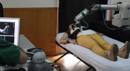
A Novel Approach for Robotic-assisted Tele-echography
Currently, robotic systems for ultrasound diagnostic procedures fall into two main categories: portable robots that require manual positioning and fully autonomous robotic systems that independently…

From Big Data to Big Decisions: How AI Stratifies Cancer Cases by Risk Factors
The CLARIFY Decision Support Platform (DSP) is a responsive web application designed to support decision-making in cancer care through real-time data integration and predictive analytics….

From “Free Text” to Structured Clinical Data: the Foundation for Clinical Decision Support Systems
Currently, the practice of recording clinical information in “free text” offers flexibility, but hinders automatic data extraction, limiting the application of analytical models. Most records…
Artificial Intelligence used in Depression Detection in Cancer Survivors
The goal of the FAITH project (Federated Artificial Intelligence solution for moniToring mental Health status after cancer treatment) is to remotely identify and predict depressive…

Integration of SONHO v2 and SClínico Systems at ULS of Coimbra to Improve Healthcare Services
With more than half a million hospital medical consultations carried out in the first half of 2024, the ULS of Coimbra stands out as an…

Elderly Care Ecosystem: an Innovative Platform for Personalized and Efficient Services
The Elderly Care Ecosystem (ECE) is an integration of various digital health technologies, exploring the areas of telehealth and predictive analytics. The goal of this…

Innovative technology that subconsciously relieves anxiety through a scarf
The SCAARF technology aims to offer an alternative method to alleviate anxiety symptoms in a non-intrusive and subconscious way. This technology is an innovative idea…

Digital Health Interventions: Equity in Hypertension Care for Everyone
Nearly half of all adults in the United States have hypertension, one of the leading risk factors for cardiovascular disease, and only about a quarter…
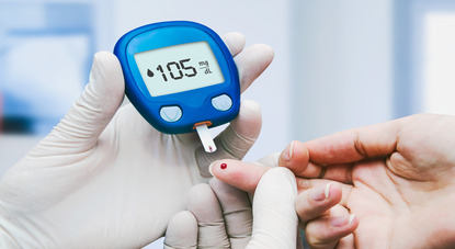
Personalization and Technology in Diabetes Management
IPDM has significant potential to improve diabetes management and drive health system reforms to become high-performing, effective, equitable, accessible, and sustainable. Evidence and good practices…

Negotiations on the European Health Data Space Advance With the Participation of the SPMS
The European Health Data Space will be a common health data sharing system across the European Union. It foresees the use of data for purposes…

Secretary of State Margarida Tavares Emphasizes Digital Innovation in Health Promotion
Margarida Tavares spoke at the opening of the conference ” O Digital na promoção contínua da saúde e do bem-estar”, organized by the Associação para…
ARS Algarve Modernizes Radiology With AI and New Data Center
The radiology service of ARS Algarve has already performed nearly 29,000 exams using Artificial Intelligence (AI) technology. In recent years, there has been a significant…

European Health Data Space: Unified Access To Health Data In The EU
The COVID-19 pandemic highlighted the importance of digital services in health, but complex rules and increasing cyberattacks make it difficult to share data across Member…

European Commission Amends Digital Europe Programme With an Investment of €762.7 Million
The European Commission has amended the Digital Europe Programme work programmes 2023-2024, investing an additional €762.7 million in Europe’s digital transition and cybersecurity. The digital…
SPMS Integrates the TEF-Health Initiative
SPMS participates in the TEF-Health initiative as a partner in a consortium composed of 51 entities from 9 European Union countries. This action is co-financed…

FMUP Creates Inhealth Junior Academy for High School Students
The InHealth Junior Academy — Academia Júnior de Inovação em Saúde is an initiative of the Departamento de Medicina da Comunidade, Informação e Decisão em…

SPMS Represents Portugal as Vice-president of GDHP
The GDHP is an intergovernmental organization in the digital health sector that facilitates cooperation and collaboration between government representatives and the World Health Organization (WHO)….
Digital Transformation of Health at INCoDe.2030 in Tomar
The “National Digital Skills Initiative e.2030, Portugal” (INCoDe.2030) is an initiative that aims to improve the Portuguese population’s level of digital skills, placing Portugal at…
Braga Hospital Evaluates Memory With Interactive Game in Patients With Multiple Sclerosis
Multiple Sclerosis is known as a chronic disease of the central nervous system, with a wide variety of motor and sensory symptoms that can lead…

Neurosurgery Teleconsultation Wins Innovation Award
The aim of the BI Award for Innovation in Healthcare is to recognize innovative projects in the healthcare sector that improve the quality of life…

HealthData@PT: New SPMS Initiative for Health Data
Action HealthData@PT is launched in the context of the implementation of the European Health Data Space, and is an initiative approved by the European Commission…
Do you have an innovative idea in healthcare field?
Share it with us and see it come to life.
We will help bring your projects to life!


Home / Publications / Publication
AUTOMATIC SEGMENTATION OF BLOOD VESSELS IN CAROTID ULTRASOUND IMAGES
Publication type: Article Summary
Original title: Vessel Detection In Carotid Ultrasound Images Using Artificial Neural Networks
Article publication date: July 2018
Source: Proceedings of the 6th International Conference Integrity, Reliability and Failure
Authors: Catarina F. Castro, Carlos C. António & Luísa C. Sousa
What is the goal, target audience, and areas of digital health it addresses?
The study aims to automatically identify blood vessels and segment carotid ultrasound images, improving diagnostic accuracy and clinical efficiency. The target audience includes healthcare professionals, such as radiologists and angiologists, as well as healthcare institutions and researchers in the fields of Artificial Intelligence (AI) and medical image processing. The study falls within the areas of digital health, with a focus on the application of AI and diagnostic support technologies.
What is the context?
Vascular diseases, such as carotid stenosis (narrowing of the carotid arteries, which connect the heart to the brain, caused by the accumulation of fatty atheroma plaques), cerebrovascular accidents (CVA) (sudden interruption of blood flow to the brain), atherosclerosis (accumulation of atheroma plaques in the arteries, leading to arterial hardening and narrowing), deep vein thrombosis (formation of blood clots in deep veins), and cervical arterial dissections (tears in the inner layer of the neck arteries), represent a significant challenge to global public health due to their high prevalence and clinical impact. Carotid stenosis is a major risk factor for CVA, affecting millions of people annually. Atherosclerosis, the leading cause of cardiovascular complications, is increasing with population ageing and risk factors such as obesity, sedentary lifestyles, and hypertension. Deep vein thrombosis, which occurs in 1 to 2 individuals per 1000 annually, can lead to pulmonary embolism (blockage of a pulmonary artery by an embolus), a severe and often fatal complication. Although less common, cervical arterial dissections are a significant cause of CVA in young adults and are frequently underdiagnosed until severe complications arise.
These conditions highlight the urgent need for advanced diagnostic tools that enable early detection and accurate assessment of these pathologies, contributing to improved clinical management and reduced complications.
What are the current approaches?
Current approaches for the diagnosis and monitoring of vascular diseases integrate advanced imaging techniques, computational methods, and clinical tools to enhance accuracy and efficiency in diagnostic outcomes. Doppler ultrasound is widely used due to its non-invasive nature, speed, and low cost, proving effective in assessing blood flow and detecting arterial obstructions. More sophisticated methods, such as computed tomography angiography and magnetic resonance angiography, provide high-resolution imaging that facilitates the detection of atheroma plaques, arterial dissections, and vascular occlusions (blockages in blood vessels). Additionally, transcranial Doppler ultrasound plays a crucial role in real-time monitoring of cerebral blood flow, proving useful in assessing conditions such as intracranial stenosis (narrowing of arteries within the skull) and other cerebrovascular disorders.
Despite their clinical utility, these techniques present limitations, including operator-dependent settings, image irregularities, and challenges in distinguishing vascular from non-vascular structures in transverse images. These factors can compromise diagnostic accuracy, particularly in cases with image artifacts or noise. Such limitations highlight the urgent need for automated, more robust, and precise solutions that can minimize variability and errors associated with manual analysis, ensuring greater consistency and accuracy in the diagnosis and monitoring of vascular diseases.
What does innovation consist of?
The innovation of this study lies in the development of an advanced methodology for the automatic segmentation of blood vessels in transverse ultrasound images of the carotid artery. The proposed approach structured into three main stages: preprocessing, geometric modelling, and classification based on artificial neural networks.
In the preprocessing stage, the region of interest in the images is binarized, removing noise smaller than 1% of the total pixels. The identification of the vascular lumen (the internal space of blood vessels) is optimized by maximizing three parameters: the circularity index, which evaluates the circular shape of structures; the irregularity index, which penalizes irregular contours; and the centrality index, which prioritizes structures positioned at the centre of the image. This combination enables a robust segmentation, accurately distinguishing the lumen from the vessel wall and assigning greyscale levels to highlight the regions of interest.
In the geometric modelling stage, Bézier curves used to smooth vascular contours. The contour pixels are organized into upper and lower subsets based on their vertical orientation, allowing for the creation of two Bézier curves that then merged to form a continuous and precise outline of the vessel walls.
In the classification stage, AI plays a key role. An advanced neural network is applied to analyse candidate regions for blood vessels, determining with high accuracy the presence or absence of vascular structures. This network was trained and tested using real ultrasound images of varying quality and acquisition settings, ensuring the model’s robustness and generalization capability across different clinical scenarios. During training, dynamic adjustments to the neural network’s internal parameters enhanced precision and minimized classification errors. Additionally, data augmentation techniques, such as horizontal image flipping, were employed to expand the dataset, significantly increasing the volume of information available for model training and validation.
What are the main results? What is the future of this approach?
Although this study does not present detailed quantitative data, the results obtained from training the model with real ultrasound images demonstrate significant advances in the accuracy and efficiency of vascular disease diagnosis. The integration of artificial neural networks in this approach enables the automated classification of candidate regions, ensuring a reliable distinction between blood vessels and adjacent structures, even in suboptimal image quality or challenging clinical conditions. Additionally, the use of Bézier curves in geometric modelling allows for the generation of precise and continuous contours of vascular walls, enhancing the detection and delineation of vascular structures. This balance between image processing techniques and AI stands out as a robust and adaptable approach that overcomes the limitations of traditional methods, such as operator-dependent settings, image noise interference, and the difficulty in distinguishing vascular from non-vascular structures.
The future of this approach is promising for transforming vascular diagnostics. Expanding the training dataset could improve the model’s generalization, making it more robust and effective across different populations and clinical settings. Its implementation in hospital software and portable ultrasound devices could enable faster, more accessible, and real-time diagnostics, particularly in regions with limited medical resources. Furthermore, this methodology can be adapted to analyse other vessels beyond the carotid artery, such as coronary and peripheral arteries, expanding its impact on cardiovascular pathologies. When combined with other imaging modalities, such as computed tomography and magnetic resonance imaging, this approach could provide more detailed and comprehensive diagnoses. Additionally, integrating clinical data, biomarkers, and demographic information allows for personalized diagnostics, aligning with the trend toward precision medicine. This innovation optimizes clinical workflows and reinforces the relevance of AI in digital health.
Would you like to know all the details?
Autonomous Robotics System for Autism Therapy
Autism spectrum disorder is a neurodevelopmental condition with significant clinical, social and economic repercussions throughout life. According to the World Health Organization, it is estimated to affect approximately 1 in 160 children worldwide. Its origin…
Virtual Reality Exergames as a Tool for Diagnosing Eye Diseases
Eye diseases represent a growing public health challenge in Portugal, significantly compromising the population’s quality of life. The increase in their prevalence is associated with various factors, such as demographic ageing, excessive use of digital…
Improving Efficiency in the Clinical Follow-up for Covid-19 Cases With a Digital Platform
COVID-19, caused by the SARS-CoV-2 virus, is a highly contagious disease with the potential to cause serious complications, requiring the isolation of infected individuals and appropriate clinical follow-up. While severe cases require hospitalization, patients with…
The Rising Threat of Antibiotic-resistant Klebsiella in Portuguese Hospitals
Healthcare-associated infections pose a serious public health threat, as they are acquired during medical treatments or hospital stays, often leading to prolonged hospitalizations, high costs for healthcare systems and high mortality rates. Portugal has one…
Deep Neural Networks And The Future Of Early Detection Of Alzheimer’s Disease
Alzheimer’s disease is the most common form of dementia, affecting more than 55 million people globally and accounting for around 70 percent of dementia cases. In Portugal, it is estimated that 200,000 people live with…
Mobile Application to Improve Workflows in Nursing Homes
Portugal has one of the highest aging populations in the world, placing increasing pressure on elderly care services, especially in nursing homes. Healthcare professionals in these facilities are often overwhelmed due to the increasing number…
The Challenges of Data Protection in Digital Health Platforms for the Elderly
Demographic ageing poses significant challenges to healthcare systems, intensifying the pressure on infrastructures and human resources. It is estimated that by 2050 the elderly population will exceed 2 billion people, making it imperative to implement…
Facilitating Epilepsy Diagnosis With a Wireless and Wearable EEG System
Paroxysmal diseases are characterized by sudden, episodic conditions that cause temporary changes in the body. Among them, epilepsy stands out for causing synchronous and uncontrolled neuronal discharges, resulting in recurrent and unprovoked seizures. These seizures…
Impact of Robotherapy-PARO on Elderly People With Dementia in Portugal
Aging is a gradual, multifactorial and continuous process characterized by the progressive loss of biological function and degeneration associated with the onset of age-related diseases. In Portugal, the aging of the population is particularly noticeable,…
New Era of Interoperability in Healthcare Systems
The growing use of electronic health records, digital diagnostic systems and remote monitoring technologies has led to a significant increase in the volume and complexity of health data. This increase intensifies the need for continuous,…
Improvement in Breast Tumor Localization With an Image Fusion Algorithm
Breast-conserving surgery aims to remove tumors while preserving as much healthy breast tissue as possible, ensuring optimal aesthetic outcomes that are critical for a patient’s quality of life. To achieve this objective, precise location of…
Collaborative Robotics Improves Working Conditions
Workers face growing challenges in the industrial environment. Among the most critical are fatigue and inappropriate postures, often associated with repetitive tasks and working conditions that lack ergonomic suitability. These factors represent significant risks for…
The Role of Mobile Technologies in the Monitoring and Rehabilitation of Peripheral Arterial Disease
PAD is a prevalent chronic condition, affecting approximately 200 million individuals globally, characterized by obstruction of the peripheral arteries, especially in the lower extremities, due to the formation of atherosclerotic plaques, which compromise blood flow…
Incorporation of Digital Implants Into CT Images to Plan Orthopedic Surgery
Orthopedic surgery addresses conditions of the musculoskeletal system to alleviate pain, restore function, and enhance the patient’s quality of life. Its success relies on meticulous pre-operative planning that incorporates clinical data and patient-specific imaging to…
Digital Health at the Top of the National Poliempreende 2024 Results
Poliempreende is a consolidated national network for encouraging entrepreneurship in higher education in Portugal, with two decades of existence. Focused on promoting innovation, the competition has had a significant impact on the national economy, with…
Digital Solution Facilitates Interaction Between Users and Health Professionals
Many patients face difficulties scheduling medical appointments in hospital units, and, when successful, they often endure long waiting times to be attended. This situation is aggravated by problems such as the incompatibility of schedules between…
The Impact of Calm Computing Integration on the Clinical Process
In recent years, digital transformation in healthcare has played a crucial role, driven by the exponential increase in medical data. This ranges from administrative information to detailed records of diagnoses, laboratory tests, medical images and…
ULS Almada-Seixal Revolutionizes With the Region’s First Surgical Robot
In recent years, ULSAS has been gradually implementing robotic systems, reinforcing its commitment to innovation and improving healthcare. Recently, the institution acquired a state-of-the-art robotic system, developed under the concept of an ‘immersive intuitive interface’,…
Online Intervention Aims to Prevent Anxiety in the General Population
Anxiety disorders are a global problem, affecting 300 million people worldwide and placing significant pressure on individuals and healthcare systems. In Europe alone, the economic impact reached 74.380 million in 2010, with 62.2% attributed to…
Rehabilitation of Facial Paralysis Through Virtual Assistants
Facial paralysis, defined by the inability to move one or both sides of the face, has an incidence of 20 to 30 cases per 100,000 people annually. This condition often causes facial weakness, difficulties in…
Detection of Anxiety and Panic Attacks in Real Time
The growing number of people with anxiety disorders, along with increased awareness of mental health, drives the need for new technological tools that provide remote and continuous monitoring of anxiety and panic disorders. Thus, the…
A Novel Approach for Robotic-assisted Tele-echography
Currently, robotic systems for ultrasound diagnostic procedures fall into two main categories: portable robots that require manual positioning and fully autonomous robotic systems that independently control the ultrasound probe’s orientation and positioning. Portable robots rely…
From Big Data to Big Decisions: How AI Stratifies Cancer Cases by Risk Factors
The CLARIFY Decision Support Platform (DSP) is a responsive web application designed to support decision-making in cancer care through real-time data integration and predictive analytics. Built on Big Data Europe, the platform integrates a variety…
From “Free Text” to Structured Clinical Data: the Foundation for Clinical Decision Support Systems
Currently, the practice of recording clinical information in “free text” offers flexibility, but hinders automatic data extraction, limiting the application of analytical models. Most records are unstructured, and the use of non-standard abbreviations increases ambiguity,…
Artificial Intelligence used in Depression Detection in Cancer Survivors
The goal of the FAITH project (Federated Artificial Intelligence solution for moniToring mental Health status after cancer treatment) is to remotely identify and predict depressive symptoms in cancer survivors using a federated machine learning approach…
Integration of SONHO v2 and SClínico Systems at ULS of Coimbra to Improve Healthcare Services
With more than half a million hospital medical consultations carried out in the first half of 2024, the ULS of Coimbra stands out as an institution dedicated to integrated, high-quality and patient-centered healthcare, with 8…
Elderly Care Ecosystem: an Innovative Platform for Personalized and Efficient Services
The Elderly Care Ecosystem (ECE) is an integration of various digital health technologies, exploring the areas of telehealth and predictive analytics. The goal of this ecosystem is to improve the quality of life for elderly…
Innovative technology that subconsciously relieves anxiety through a scarf
The SCAARF technology aims to offer an alternative method to alleviate anxiety symptoms in a non-intrusive and subconscious way. This technology is an innovative idea in the field of digital health and wearable technology, designed…
Digital Health Interventions: Equity in Hypertension Care for Everyone
Nearly half of all adults in the United States have hypertension, one of the leading risk factors for cardiovascular disease, and only about a quarter (24%) of those people have their hypertension under control. Studies…
Personalization and Technology in Diabetes Management
IPDM has significant potential to improve diabetes management and drive health system reforms to become high-performing, effective, equitable, accessible, and sustainable. Evidence and good practices inspire health system transformation. Adopting person-centred approaches like co-creation and…
Negotiations on the European Health Data Space Advance With the Participation of the SPMS
The European Health Data Space will be a common health data sharing system across the European Union. It foresees the use of data for purposes that benefit people and society. It will ensure citizens have…
Secretary of State Margarida Tavares Emphasizes Digital Innovation in Health Promotion
Margarida Tavares spoke at the opening of the conference ” O Digital na promoção contínua da saúde e do bem-estar”, organized by the Associação para a Promoção e Desenvolvimento da Sociedade da Informação (APDSI) and…
ARS Algarve Modernizes Radiology With AI and New Data Center
The radiology service of ARS Algarve has already performed nearly 29,000 exams using Artificial Intelligence (AI) technology. In recent years, there has been a significant investment in image digitization and data storage, as well as…
European Health Data Space: Unified Access To Health Data In The EU
The COVID-19 pandemic highlighted the importance of digital services in health, but complex rules and increasing cyberattacks make it difficult to share data across Member States; the EHDS, based on several regulations, provides tailor-made rules…
European Commission Amends Digital Europe Programme With an Investment of €762.7 Million
The European Commission has amended the Digital Europe Programme work programmes 2023-2024, investing an additional €762.7 million in Europe’s digital transition and cybersecurity. The digital transition is the main work programme with a budget of…
SPMS Integrates the TEF-Health Initiative
SPMS participates in the TEF-Health initiative as a partner in a consortium composed of 51 entities from 9 European Union countries. This action is co-financed by the European Commission and has a duration of five…
FMUP Creates Inhealth Junior Academy for High School Students
The InHealth Junior Academy — Academia Júnior de Inovação em Saúde is an initiative of the Departamento de Medicina da Comunidade, Informação e Decisão em Saúde da Faculdade de Medicina da Universidade do Porto (FMUP)….
SPMS Represents Portugal as Vice-president of GDHP
The GDHP is an intergovernmental organization in the digital health sector that facilitates cooperation and collaboration between government representatives and the World Health Organization (WHO). Its purpose is to foster policymaking that promote the digitalization…
Digital Transformation of Health at INCoDe.2030 in Tomar
The “National Digital Skills Initiative e.2030, Portugal” (INCoDe.2030) is an initiative that aims to improve the Portuguese population’s level of digital skills, placing Portugal at the level of the most advanced European countries in this…
Braga Hospital Evaluates Memory With Interactive Game in Patients With Multiple Sclerosis
Multiple Sclerosis is known as a chronic disease of the central nervous system, with a wide variety of motor and sensory symptoms that can lead to work disability, socioeconomic burden, and reduced quality of life…
Neurosurgery Teleconsultation Wins Innovation Award
The aim of the BI Award for Innovation in Healthcare is to recognize innovative projects in the healthcare sector that improve the quality of life for the Portuguese people. In 2021, the specific theme was…
HealthData@PT: New SPMS Initiative for Health Data
Action HealthData@PT is launched in the context of the implementation of the European Health Data Space, and is an initiative approved by the European Commission under the EU4Health 2021-2027 programme. This initiative contributes to the…
Do you have an innovative idea in healthcare field?
Share it with us and see it come to life.
We will help bring your projects to life!